ADAS 3D
friend for the
The new imaging
electrophysiologist


Given Luis Serra’s passion for photography, it is only natural that ADAS3D Medical —a company that emerged from the Department of Information Technologies at the Universitat Pompeu Fabra in Barcelona, of which he is the president—wants to build “something like Photoshop” for the cardiac imaging space.
“Photography and cinema are two of my main hobbies,” says Serra, a PhD in electrical engineering who has over 20 years of experience in the development of medical applications powered by Virtual Reality and Augmented Reality technologies. “I remember being amazed by 3D movies when I was growing up, when they first emerged on the scene. In fact, that is where a lot of my interest in 3D medical imaging was born.”
The Barcelona-based company—ADAS stands for Automatic Detection of Arrhythmic Substrate—is focussed on developing post-processing software solutions that translate cardiac MRI and CT images into visual 3D images that can aid doctors in identifying areas that may trigger arrhythmias (a fast heart rhythm that if not treated could be lethal) and planning ablation procedures. It also has a partnership with Circle Cardiovascular Imaging, the market leader in MR and CT post-processing software, for distribution and joint development of MRI scanner application products and workflows, with corresponding post-processing capabilities.
Serra pursued his interest in 3D medical imaging first in the area of neurosurgery, when he co-founded Volume Interactions in Singapore, a company specialising in software and hardware for pre-operative planning and image-guided surgery in 2000 that was acquired by the Bracco Group (Milan, Italy) a couple of years later. “My first imaging experience was a great learning opportunity and provided a very good training ground to set me up for my next venture” said Serra.
He chanced upon the opportunity to apply these learnings to cardiology following his return to Barcelona in 2010, where he met doctors and engineers who shared similar interests at the Universitat Pompeu Fabra. They co-founded the company and worked out of the university for close to seven years, acquiring a solid footing in developing imaging post-processing software solutions by participating in national and international competitive projects and nurturing close collaborations with several clinical institutions, especially the Hospital Clinic of Barcelona.
“We were very certain that we wanted to address a gap—to cater to the imaging needs of electrophysiologist who don't have too much exposure to 3D imaging because they are trained on the electricity side. But, they need help to perform complex interventions inside the heart. That is the potential we wanted to tap” recalled Serra.
Second coming
An electrocardiogram, which detects and records the timing and strength of electrical signals as they travel through the heart, is the most common tool used to diagnose ventricular tachycardia (VT). Although doctors can determine the type of tachycardia a patient has by looking for patterns among the signals, electrocardiograms are limited when it comes to planning interventions.
The electrocardiograms can only give doctors a rough idea about the location of scars; they do not give the physician a detailed picture of to expect when they bring the patient in to the EP lab to treat them. When the patient is on the table, the physician must manipulate a catheter from the patient’s groin into the patient’s left ventricle, which is one meter away, and carefully find very specific areas to ablate. This means the physician must search for the electrical short circuits which can be time consuming and if he can’t find one, this can result in recurrence of tachycardia after ablation, which is high across patient populations.
This is where Serra says the company ADAS 3D imaging platform is “the new imaging friend for the electrophysiologist”. Its software solutions are designed to bridge the gap between radiology, cardiology, and physiology, interpreting what each has to say about the condition of the heart, and combining them to create 3D images identifying problem areas and delineating the need and scope for interventions. These 3D images not only provide information on scar quantification and location but also characterization—the scar tissue characterization is depicted through color-coded pixel signal intensity maps that “have a reasonably high correlation with the electro-anatomic maps obtained during the ablation procedures,” shows research.
“Cardiac arrhythmias can originate from many places in the heart that are difficult to quickly and easily find with available technology and techniques. Effectively, our software allows electrophysiologists to get inside the heart, to have more detailed, up-close information, to go through the myocardial walls…represented in 3D images, ahead of time. This allows them to know what to expect and to plan where they may need to look closer during the procedure. This is something that is new and exciting for the field.” says Serra.
The new imaging friend for the electrophysiologist
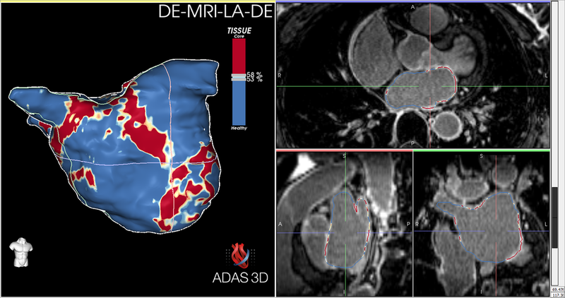
ADAS 3D currently offers two products—ADAS 3D LA, designed to reveal fibrosis in the atria; and ADAS 3D LV, designed to identify arrhythmia substrate and plan ablation procedures. ADAS 3D LA provides electrophysiologists with tools to define the shape of left atrium from Late Gadolinium Enhancement (LGE) 3D MRI data. It then automatically computes the IIR value of the atrial wall and quantifies the fibrosis, the atrial volume and the sphericity. It also allows exporting the segmented left atrium 3D shape and associated fibrosis into formats compatible with EP navigation systems.
ADAS 3D LV addresses the catheter ablation treatment needs of electrophysiologists, before, during and after interventions. At the pre-intervention stage, the application allows electrophysiologists to use MRI data of a patient with VT to obtain the left ventricle, right ventricle and aorta, and to detect potential 3D Corridors in the left ventricle, represented by different colours. This planning data, including the 3D Corridors and other relevant myocardial information, comes in handy during interventions as it can be exported from ADAS 3D LV to files in formats accepted by EP navigation system. These files can be imported into the catheter navigation system and used to assist the electro-anatomical mapping and the ablation. The application can also load data from the EP navigation system after the intervention, which can be used to compare MRI information to electro-anatomical data. Studies show that the applications significantly reduce chances of relapse of tachycardia and the need for subsequent ablation procedures.
Like using any new tool such as Word or Photoshop, there is a short learning curve to learn how to use the ADAS 3D software and apply it to diagnosing and treating patients. Hospitals that buy the ADAS 3D software licence can use it for all their complex cases without worrying about per case subscription fees and without transmitting the data via the cloud for processing that can cause patient confidentiality concerns.
At present, the interpretation of MRI and CT images is done semi-automatically, and the process takes about 10-15 minutes. The team at ADAS3D Medical, in collaboration with Circle Cardiovascular Imaging, is working on reducing the window by incorporating deep learning and machine learning technologies, and providing better integration between CT and MRI.
Moving forward, the company also wants to augment its offerings by expanding into all the different chambers of the heart. “To date, the thin walled structures like the right ventricle and the right atria have been difficult to image and thus study at the imaging level… so we have our work cut out.” says Serra. The company is also working on offering its applications on web browsers, and enabling electrophysiologists to not just visualize but also interact with 3D images using immersive, interactive stereoscopic monitors.
Custom tools
Road ahead
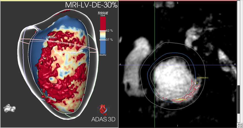

The increasing availability of high-quality patient images enables ADAS 3D's algorithms and software to extract information to support clinical decision-making and planning and guidance of interventional procedures. Using ADAS 3D products, electrophysiologists can identify the arrhythmic substrate and plan catheter ablation procedures.
By Michelle thoeny
My first imaging experience was a great learning opportunity and provided a very good training ground to set me up for my next venture
Dr Luis Serra, Director ADAS 3D